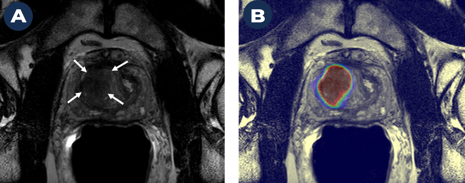
We evaluated a patient with a high PSA level (indicating a risk for prostate cancer) and a strong family history of prostate cancer. A Magnetic Resonance Image (MRI) on the left (A) shows a suspicious cancer lesion (marked by arrows). The image on the right (B) shows a high probability of prostate cancer within this lesion based on a deep learning-based AI algorithm. We biopsied the lesion using a technology that “fuses” transrectal ultrasound with MRI and were able to identify the early stages of clinically significant prostate cancer (Gleason 3+4).