News
Using Artificial Intelligence to Map Prostate Tumors
In a retrospective study, NCI-funded researchers examined the usefulness of artificial intelligence (AI) in mapping prostate cancer.
They used an AI-driven approach that blended biopsy and clinical results with Magnetic Resonance Imaging (MRI) to see if this model could predict tumor margins better than using MRI alone.
Being able to accurately delineate tumor margins is key, as it allows oncologists to precisely target treatment to the cancer tumor (i.e., referred to as focal therapy). Defining margins using MRI has been the conventional approach, but MRI is known to underestimate tumor margins.
The researchers turned to AI in an effort to map a more complete picture of the tumor. The study’s corresponding author, Dr. Alan Priester from the David Geffen School of Medicine, University of California-Los Angeles, noted, “When AI is used to analyze multimodal information—MRI, tracked biopsy findings, and PSA—tumor delineation can be greatly improved.”
The investigators used an AI model (“Unfold AI”) developed by Avenda Health and cleared by the U.S. Food and Drug Administration. They trained the model using tracked biopsy data, clinical information, and histopathology results, and then they tested it using an independent data set.
Using this AI model, the researchers were able to develop an accurate cancer estimation map and define more effective tumor margins. They found their model performed better than conventional estimates of tumor margins using MRI alone.
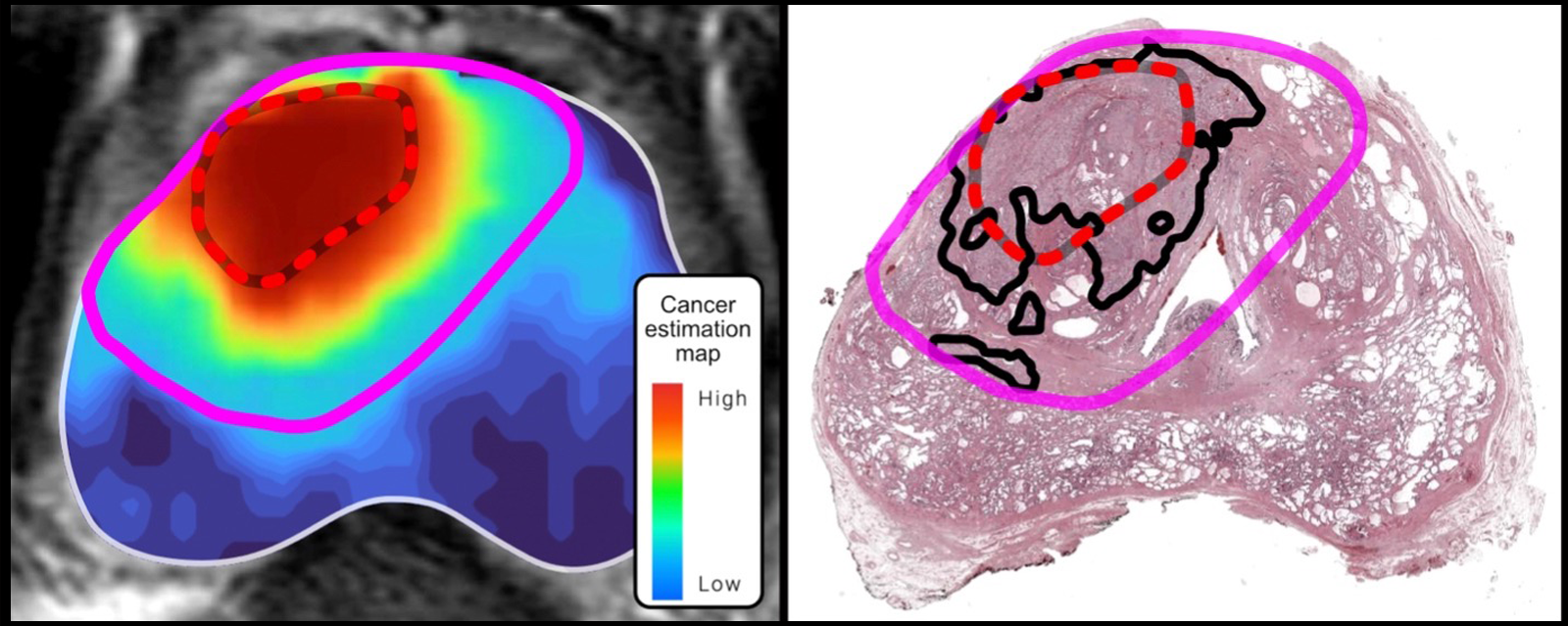
Cancer estimation map (left image) shows the difference between an AI-approach to defining the tumor margins (pink line) and a conventional approach that relies on MRI alone (red-dotted line). In the AI model, the tumor margin extends well beyond the margins identified using a conventional approach. Histopathology (right image) confirms that the AI model successfully enclosed the full extent of the tumor (black line).
Dr. Priester said, “In our study, treatment of the original MRI-visible tumor would have resulted in positive margins for every patient, which means targeted therapy would have missed cancer outside the region of interest. By contrast, AI margins were negative 80–90 percent of the time, which means targeted therapy would have succeeded.”
The study authors acknowledge some study limitations, including the fact that data were from one institution and featured a population that likely had more advanced disease than the typical focal therapy patient.
In future studies, the researchers plan to apply the model to broader populations and to incorporate diffusion-weighted MRI, which should give an even more complete cancer estimation map than the images used in the current study.